Written by: Jorie Larsen Schroeder
Reviewed by: Nina Kalus, PsyD, and Yalda Safai, MD, MPH
TMS is a noninvasive, FDA-cleared neuromodulation therapy for treating major depressive disorder (MDD), obsessive-compulsive disorder (OCD), and other mental health conditions. It works by generating a magnetic field that induces an electric current in the brain, activating neurons in targeted regions.
The type of coil matters. Different TMS coil configurations produce different electric field distributions, changing how the therapy reaches the brain.
TMS has been shown to improve neuroplasticity and increase cortical excitability, though researchers are still working out its specific mechanisms of action.
TMS is cumulative, not a one-off intervention. Symptoms typically improve gradually over four to six weeks of consistent treatment.
Transcranial magnetic stimulation (TMS) is an FDA-cleared, noninvasive neuromodulation therapy that directly stimulates brain structures associated with mental health conditions including major depressive disorder (MDD), obsessive-compulsive disorder (OCD), and more.
If your patients are asking “How does TMS work?”, continue reading for a clinical explanation as we explore the actual mechanism of action behind TMS: how a magnetic pulse goes on to trigger a change in neuronal activity—and most importantly, what that means for your patients’ treatment plan.
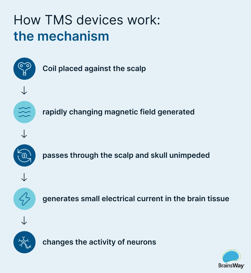
TMS therapy involves the use of an electromagnetic coil placed against the scalp, which generates brief pulses of magnetic energy. The magnetic field passes through the scalp and skull and induces an electric current in the underlying brain tissue. From there, it can stimulate neurons and bolster neuroplasticity, or the brain’s ability to change and adapt by forming new connections between nerve cells.
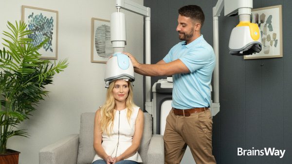
During a typical TMS treatment session, a patient sits in a comfortable treatment chair and the provider positions a coil against the patient’s scalp. After completing the motor threshold check, which helps determine the proper placement on the skull for each individual patient, the provider calibrates the individual stimulation settings. A typical course of treatment is one 20-minute session, five days a week, over a period of four to six weeks, for a total of about 30 to 36 sessions.
TMS itself is structured around the basic scientific principles discovered by English physicist and chemist Michael Faraday in 1831, proving that magnetic fields can induce electrical currents. TMS as a neuromodulation therapy began in the 1980s.
Although TMS is a noninvasive outpatient procedure that typically takes about 20 minutes per session, there is much more happening beneath the surface. If you were to slow down each magnetic pulse and follow it into the brain to study its effect, here’s what you’d see.
TMS devices generate highly concentrated magnetic fields, which turn on and off very rapidly. These magnetic fields—the same type and strength as those created by a magnetic resonance imaging (MRI) machine—pass through the scalp and skull unimpeded, inducing an electrical current in the cortex beneath it.
It’s also important to note: Because TMS produces strong, pulsed magnetic fields, certain metallic and electronic implants can pose a patient safety risk. That’s why TMS is contraindicated in patients with implanted devices in the head or neck, including cochlear implants.
Next, once the electric current is induced within the brain, it activates the neurons within the affected cortical tissue.
“Inside of the brain, neurons communicate with each other via both electrical and chemical (neurotransmitter) signals,” explains Ben Spielberg, MS, PhD candidate, and founder and CEO at Solstice Training Institute.
Dendrites, a branch-like part of a neuron that acts like an antenna, receive signals from other neurons, producing changes in the neuron’s membrane potential. At the axon terminal, that electrical signal can trigger the release of neurotransmitters into the synaptic cleft, in turn influencing the activity of the next neuron in the chain.
Spielberg explained that this process allows signals initiated in one area of the brain to travel down connected pathways, influencing broader brain networks.
When treating major depressive disorder, providers typically target the left dorsolateral prefrontal cortex, an area of the brain associated with mood regulation. The magnetic pulses stimulate these specific neurons, which are in turn connected via an intricate circuit with deeper brain structures that regulate emotions and process sadness.
“The neurons we stimulate are connected to many other areas of the brain like a ripple effect or ocean wave,” says Vinay Saranga, MD, psychiatrist at the North Carolina Institute of Advanced NeuroHealth. “So when we activate neurons in one cortical region, that activity can spread through those existing connections and influence other parts of the brain.”
He continues: “The really neat thing about TMS I think isn’t really treating only the small piece of cortex underneath the coil. Again, we’re using that cortical area as an access point to influence a much larger, interconnected brain network.”
Spielberg offers another analogy.
“Think of a neural network like a subway system, with each node being a stop on the subway,” says Spielberg. “There are different subway lines to get you to different places,” just as there are different neural networks connecting different regions of the brain.
“With TMS for depression in particular,” adds Spielberg, “we generally stimulate the left dorsolateral prefrontal cortex hoping that the signal will propagate to the subgenual anterior cingulate cortex, which is a key target in depression treatment.”
Some of these stimulated neurons go on to release increased levels of neurotransmitters that play an important role in mood stabilization, including serotonin, dopamine, and norepinephrine. But saying depression is caused only by an imbalance of neurotransmitters grossly oversimplifies the disease.
The way we’ve been thinking about depression over the years doesn’t match what the neuroscience of depression tells us, according to Spielberg. “Depression isn’t necessarily a neurotransmitter imbalance, but rather a consequence of deficits in functional connectivity between different parts of the brain,” he explains. “TMS is unique in the ability to reorganize the brain’s functional connectivity without the negative side effect profile of oral antidepressant medication.”
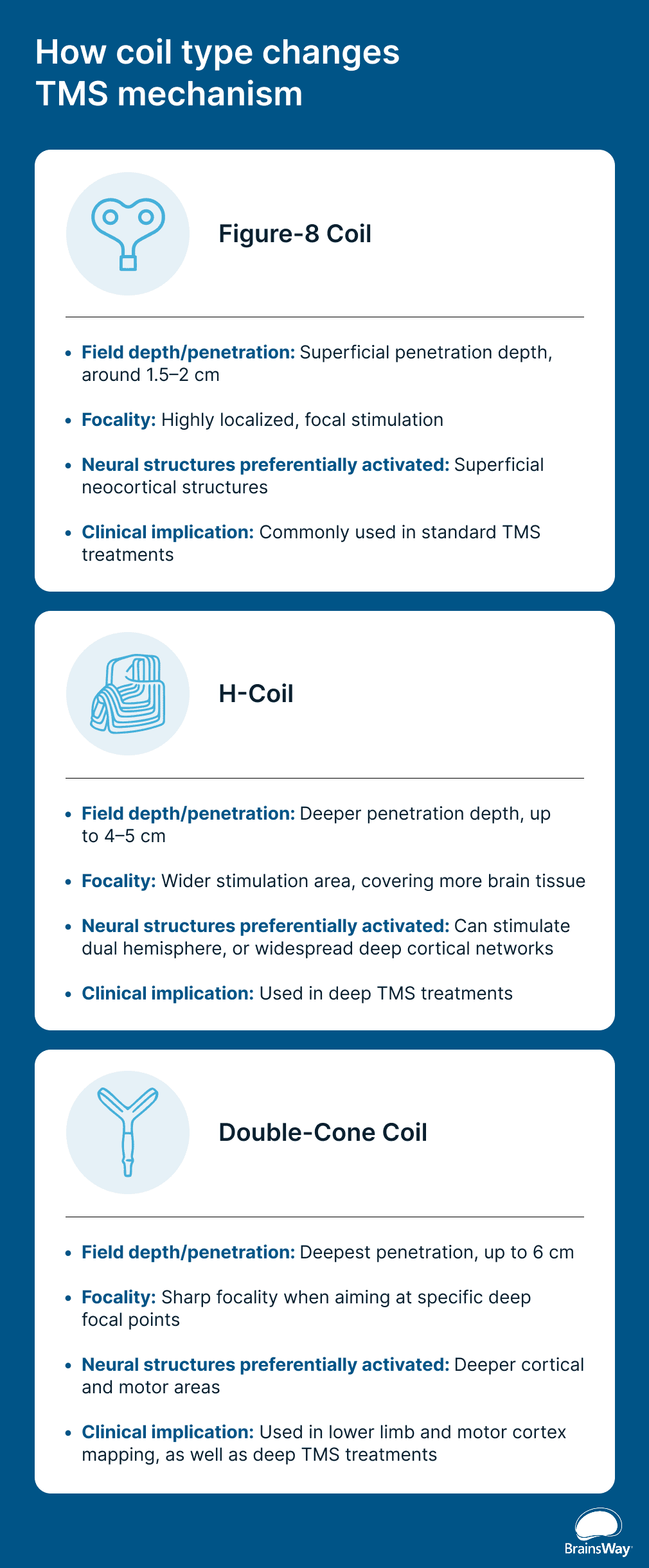
The type of coil used can have significant impacts on how TMS therapy works, as different TMS coil configurations produce different electric field distributions.
The earliest TMS devices dating back to the 1980s used a circular coil; in 2008, the figure-8 coil became the first FDA-cleared TMS coil for depression. The figure-8 coil is still widely used in standard TMS and known for its highly focal, yet more superficial, stimulation.
In 2013, the BrainsWay-patented H-Coil—designed to deliver deep TMS (dTMS) to depths up to 5 centimeters within the brain’s deeper structures—was FDA-cleared to treat major depressive disorder.
“With standard TMS, we can be very precise about where we’re stimulating, but the stimulation is mostly limited to the outer layers of the brain, or the cortex,” says Dr. Saranga.
Deep TMS reaches deeper, and while it’s technically less focused, it stimulates a wider area. “Even with deep TMS, we’re not simply reaching down and directly stimulating one specific deep brain structure. We’re still stimulating a broader network of brain tissue,” says Dr. Saranga.
The double-cone coil can penetrate up to 6 centimeters within the brain tissue, making it ideal for deep TMS as well as lower limb and motor cortex mapping.
| Type of Coil | Figure-8 Coil | H-Coil | Double-Cone Coil |
|---|---|---|---|
| Field depth/penetration | Superficial penetration depth, around 1.5–2 cm | Deeper penetration depth, up to 4–5 cm | Deepest penetration, up to 6 cm |
| Focality | Highly localized, focal stimulation | Wider stimulation area, covering more brain tissue | Sharp focality when aiming at specific deep focal points |
| Neural structures preferentially activated | Superficial neocortical structures | Can stimulate dual hemisphere, or widespread deep cortical networks | Deeper cortical and motor areas |
| Clinical implication | Commonly used in standard TMS treatments | Used in BrainsWay’s patented deep TMS treatments | Used in lower limb and motor cortex mapping, as well as deep TMS treatments |
Coil type isn’t the only variable. Providers can also adjust the frequency and pattern of stimulation depending on the desired outcome. Here’s a quick breakdown.
While the efficacy of TMS in treating major depressive disorder, obsessive-compulsive disorder (OCD), and more is well-documented, there is active research underway to determine the specific mechanisms of action that drive its therapeutic effects.
David Wolff, MD, MPH, and medical director of New Life Mental Health, says that the front-end process of TMS is fairly clearly established, wherein the fluctuating electric current changes the pattern of neuron firing. “This increases the excitability of the cortex being stimulated, and likely changes the patterns of communication with adjacent brain regions,” he says.
However, Dr. Wolff advises providers to avoid using overly simplistic terms when describing TMS’ effects, noting that phrases like “TMS turns on an underactive portion of the brain” or “permanently rewires the brain” don’t tell the full story.
He says that both of these are effective ways to describe what the patient may feel has occurred, but from a biological perspective, research cannot yet support the specific mechanisms and cellular changes involved.
It’s important to explain to patients that TMS is not a one-time solution. Over the course of four to six weeks of consistent treatment, changes are often gradual and cumulative as neuroplasticity effects begin to take shape. Progress is not always linear, but many patients report that their symptoms are improving after just a few weeks.
“An additional analogy to help understand how TMS produces its long-term effects could be drawn to physical exercise,” says Dr. Wolff. “While a single workout produces acute physiological effects, repeated workouts can cause gradual lasting changes in how your body operates.”
TMS works through a physiological chain that scientists understand increasingly well: an electromagnetic coil generates a magnetic field, which activates neurons, which boosts neuroplasticity and cortical excitability. As Dr. Saranga says, “Depression is not just caused by dysfunction in one isolated region of the brain. It involves distributed networks. With TMS, we can hit these different regions in a controlled and repetitive manner which helps us reduce or alleviate a patient’s symptoms.”
For providers, tailoring a TMS treatment plan means understanding how coil design, stimulation target, intensity, and frequency influence where and how the induced electrical field interacts with the brain.
During transcranial magnetic stimulation (TMS) therapy, an electromagnetic coil positioned against the scalp generates brief pulses of magnetic energy. The magnetic field passes through the skull and induces an electric current in the underlying brain tissue, stimulating neurons. Providers can choose from a wide range of TMS devices, coil types, frequencies, positions on the scalp, and more to tailor the treatment to a specific patient and their condition.
Yes, TMS (transcranial magnetic stimulation), causes neurons to activate, which in turn can cause them to release neurotransmitters including dopamine, serotonin, and norepinephrine.
TMS (transcranial magnetic stimulation) therapy targets the left dorsolateral prefrontal cortex in order to stimulate neurons in this brain region, which is associated with mood regulation. These neurons are connected to complex structures deeper in the brain that further regulate emotion.
TMS (transcranial magnetic stimulation) is a cumulative therapy, not a single intervention. Patients should expect gradual changes over the course of several weeks of consistent treatment as their brain’s networks begin functioning more efficiently.